

 Book an Appointment
 Patient Portal
Shoulder
Shoulder Arthroscopy
Arthroscopy is a minimally invasive diagnostic and surgical procedure performed for joint problems. Shoulder arthroscopy is performed using a pencil-sized instrument called an arthroscope. The arthroscope consists of a light system and camera that projects images of the surgical site onto a computer screen for your doctor to clearly view.
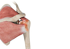
Shoulder Reconstruction Surgery
Shoulder reconstruction surgery is an operative procedure in which stretched or torn soft-tissue structures that surround the shoulder joint such as the capsule, ligaments, and cartilage, are repaired to secure the shoulder joint in place.
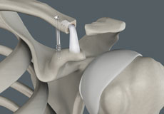
Acromioclavicular Joint Reconstruction
Shoulder separation can usually be managed by non-surgical treatments. In cases of a severe separation of the AC joint, your surgeon may perform a surgical repair or use a tissue graft to reconstruct the damaged ligaments.
Rotator Cuff Repair
Rotator cuff repair is a surgery to repair an injured or torn rotator cuff. It is usually performed arthroscopically on an outpatient basis. An arthroscope, a small, fiber-optic instrument consisting of a lens, light source, and video camera.
SLAP Repair
A SLAP repair is an arthroscopic shoulder procedure to treat a specific type of injury to the labrum called a SLAP tear.
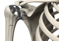
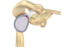
Shoulder Joint Replacement
Total shoulder replacement surgery is performed to relieve symptoms of severe shoulder pain and disability due to arthritis. In this surgery, the damaged articulating parts of the shoulder joint are removed and replaced with artificial prostheses. Replacement of both the humeral head and the socket is called a total shoulder replacement.
Subacromial Decompression
Subacromial decompression is a surgical procedure performed for the treatment of a condition called shoulder impingement. In shoulder impingement, the degree of space between the rotator cuff tendon and shoulder blade is decreased due to irritation and swelling of the bursa or due to development of bone spurs.
Reverse Total Shoulder Replacement
Conventional surgical methods such as total shoulder joint replacement are not very effective in the treatment of rotator cuff arthropathy. Reverse total shoulder replacement is an advanced surgical technique specifically designed for rotator cuff tear arthropathy, a condition where you suffer from both shoulder arthritis and a rotator cuff tear.
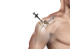
Shoulder Fracture Care
A break in the bone that makes up the shoulder joint is called a shoulder fracture. The clavicle (collarbone) and end of the humerus (upper arm bone) closest to the shoulder are the bones that usually are fractured. The scapula, or shoulder blade, is not easily fractured because of its protective cover of surrounding muscles and chest tissue.
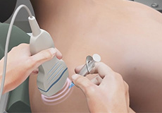
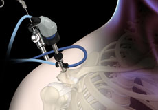
Ultrasound Guided Shoulder Injections
An ultrasound-guided injection is a minimally invasive procedure used for Treating various painful musculoskeletal conditions such as tendonitis, bursitis, and neuritis. Performing cyst aspiration Guiding the placement of needles for both diagnostic as well as therapeutic purposes
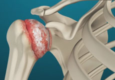
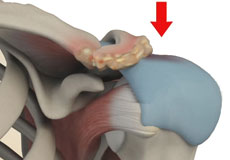
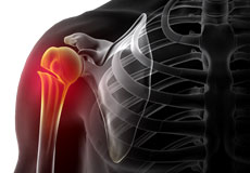
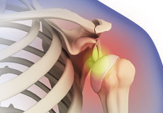
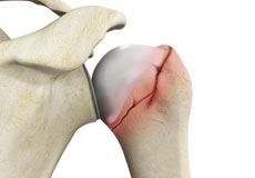
Shoulder Arthritis
The term arthritis literally means inflammation of a joint but is generally used to describe any condition in which there is damage to the cartilage. Damage of the cartilage in the shoulder joint causes shoulder arthritis. Inflammation is the body's natural response to injury. The warning signs that inflammation presents are redness, swelling, heat, and pain.
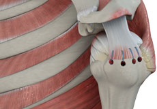
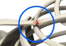
Rotator Cuff Tear
A rotator cuff is a group of tendons in the shoulder joint that provides support and enables a wide range of motion. A major injury to these tendons may result in rotator cuff tears. It is one of the most common causes of shoulder pain in middle-aged and older individuals.
Shoulder Instability
Shoulder instability is a chronic condition that causes frequent dislocation of the shoulder joint.
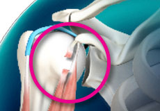
Shoulder Impingement
Shoulder impingement is the inflammation of the tendons of the shoulder joint. It is one of the most common causes of pain in the shoulder. Shoulder impingement is also called swimmer’s shoulder, tennis shoulder or rotator cuff tendinitis.
SLAP Tears
The term SLAP (superior –labrum anterior-posterior) lesion or SLAP tear refers to an injury of the superior labrum of the shoulder.
Frozen Shoulder
Frozen shoulder, also called adhesive capsulitis, is a condition in which you experience pain and stiffness in your shoulder. The symptoms appear slowly, worsen gradually and usually take one to three years to resolve on their own.
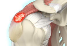
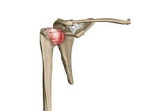
Calcification Tendonitis of the Shoulder
Calcification tendinitis is a problem with the shoulder’s tendons and muscles. This condition occurs due to the formation of calcium deposits in the tendons (tissue which attaches muscle to bone) of the rotator cuff (a group of muscles and tendons stabilizing the shoulder). This calcium build-up causes inflammation of the tissues surrounding it, and intense shoulder pain.
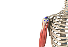
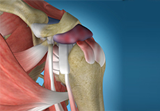
Long Head Biceps Tendon Rupture
Your biceps muscle has two heads, a long head, and a short head, which are both attached to the shoulder. The long head of the biceps tendon is a tough band of connective fibrous tissue that attaches the long head of the biceps to the top of the shoulder socket.
Shoulder Dislocation
Sports that involve overhead movements and repeated use of the shoulder at your workplace may lead to sliding of the upper arm bone from the glenoid. The dislocation might be a partial dislocation (subluxation) or a complete dislocation causing pain and shoulder joint instability.
Bicep Tendon Rupture at Shoulder
Overuse and injury can cause fraying of the biceps tendon and eventual rupture. A biceps tendon rupture can either be partial, where it does not completely tear the tendon or complete, where the tendon completely splits in two and is torn away from the bone.
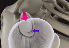
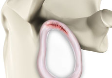
Shoulder Labrum Tear
Traumatic injury to the shoulder or overuse of the shoulder (throwing, weightlifting) may cause the labrum to tear. In addition, aging may weaken the labrum leading to injury.
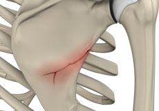
Shoulder Fractures
A break in a bone that makes up the shoulder joint is called a shoulder fracture.
Clavicle Fracture (Broken Collarbone)
The break or fracture of the clavicle (collarbone) is a common sports injury associated with contact sports such as football and martial arts, as well as impact sports such as motor racing. A direct blow over the shoulder that may occur during a fall on an outstretched arm or a motor vehicle accident may cause the clavicle bone to break.
Fracture of the Shoulder Blade
The scapula (shoulder blade) is a flat, triangular bone providing attachment to the muscles of the back, neck, chest and arm. The scapula has a body, neck and spine portion.
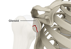
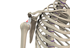
Glenoid Fracture
Fractures of the glenoid are rare but can occur due to major trauma or during high-energy sports activities.
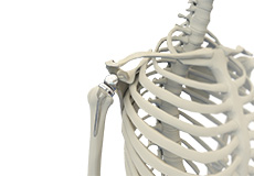
Shoulder Anatomy
The shoulder is the most flexible joint in the body that enables a wide range of movements including forward flexion, abduction, adduction, external rotation, internal rotation, and 360-degree circumduction. Thus, the shoulder joint is considered the most insecure joint of the body, but the support of ligaments, muscles, and tendons function to provide the required stability.
Bones of the Shoulder
The shoulder joint is a ball and socket joint made up of three bones, namely the humerus, scapula, and clavicle.
Humerus
The end of the humerus or upper arm bone forms the ball of the shoulder joint. An irregular shallow cavity in the scapula called the glenoid cavity forms the socket for the head of the humerus to fit in. The two bones together form the glenohumeral joint, which is the main joint of the shoulder.
Scapula and Clavicle
The scapula is a flat triangular-shaped bone that forms the shoulder blade. It serves as the site of attachment for most of the muscles that provide movement and stability to the joint. The scapula has four bony processes - acromion, spine, coracoid and glenoid cavity. The acromion and coracoid process serve as places for attachment of the ligaments and tendons.
The clavicle bone or collarbone is an S-shaped bone that connects the scapula to the sternum or breastbone. It forms two joints: the acromioclavicular joint, where it articulates with the acromion process of the scapula and the sternoclavicular joint where it articulates with the sternum or breast bone. The clavicle also forms a protective covering for important nerves and blood vessels that pass under it from the spine to the arms.
Soft Tissues of the Shoulder
The ends of all articulating bones are covered by smooth tissue called articular cartilage, which allows the bones to slide over each other without friction, enabling smooth movement. Articular cartilage reduces pressure and acts as a shock absorber during movement of the shoulder bones. Extra stability to the glenohumeral joint is provided by the glenoid labrum, a ring of fibrous cartilage that surrounds the glenoid cavity. The glenoid labrum increases the depth and surface area of the glenoid cavity to provide a more secure fit for the half-spherical head of the humerus.
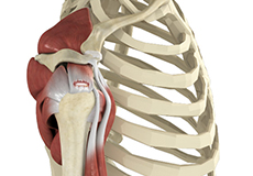
Ligaments of the Shoulder
Ligaments are thick strands of fibers that connect one bone to another. The ligaments of the shoulder joint include:
- Coracoclavicular ligaments: These ligaments connect the collarbone to the shoulder blade at the coracoid process.
- Acromioclavicular ligament: This connects the collarbone to the shoulder blade at the acromion process.
- Coracoacromial ligament: It connects the acromion process to the coracoid process.
- Glenohumeral ligaments: A group of 3 ligaments that form a capsule around the shoulder joint and connect the head of the arm bone to the glenoid cavity of the shoulder blade. The capsule forms a watertight sac around the joint. Glenohumeral ligaments play a very important role in providing stability to the otherwise unstable shoulder joint by preventing dislocation.
Muscles of the Shoulder
The rotator cuff is the main group of muscles in the shoulder joint and is comprised of 4 muscles. The rotator cuff forms a sleeve around the humeral head and glenoid cavity, providing additional stability to the shoulder joint while enabling a wide range of mobility. The deltoid muscle forms the outer layer of the rotator cuff and is the largest and strongest muscle of the shoulder joint.
Tendons of the Shoulder
Tendons are strong tissues that join muscle to bone allowing the muscle to control the movement of the bone or joint. Two important groups of tendons in the shoulder joint are the biceps tendons and rotator cuff tendons.
Bicep tendons are the two tendons that join the bicep muscle of the upper arm to the shoulder. They are referred to as the long head and short head of the bicep.
Rotator cuff tendons are a group of four tendons that join the head of the humerus to the deeper muscles of the rotator cuff. These tendons provide more stability and mobility to the shoulder joint.
Nerves of the Shoulder
Nerves carry messages from the brain to muscles to direct movement (motor nerves) and send information about different sensations such as touch, temperature, and pain from the muscles back to the brain (sensory nerves). The nerves of the arm pass through the shoulder joint from the neck. These nerves form a bundle at the region of the shoulder called the brachial plexus. The main nerves of the brachial plexus are the musculocutaneous, axillary, radial, ulnar and median nerves.
Blood vessels of the Shoulder
Blood vessels travel along with the nerves to supply blood to the arms. Oxygenated blood is supplied to the shoulder region by the subclavian artery that runs below the collarbone. As it enters the region of the armpit, it is called the axillary artery and further down the arm, it is called the brachial artery.
The main veins carrying de-oxygenated blood back to the heart for purification include:
- Axillary vein: This vein drains into the subclavian vein.
- Cephalic vein: This vein is found in the upper arm and branches at the elbow into the forearm region. It drains into the axillary vein.
- Basilic vein: This vein runs opposite the cephalic vein, near the triceps muscle. It drains into the axillary vein.
Elbow
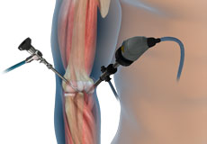
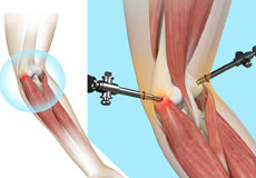
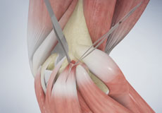
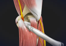
Elbow Arthroscopy
Elbow arthroscopy, also referred to as keyhole or minimally invasive surgery, is a surgical procedure that is performed through tiny incisions to evaluate and treat several elbow conditions.
Tennis Elbow Surgery
Tennis elbow is a painful condition occurring from repeated muscle contractions at the forearm. The condition is more common in sports activities such as tennis, painting, hammering, typing, gardening and playing musical instruments.
Golfer's Elbow Surgery
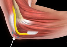
Golfer’s elbow is a condition associated with pain on the inside of the elbow where tendons of your forearm attach to the bony prominence (medial epicondyle). It is also called medial epicondylitis and is caused by injury or irritation to the tendons which can become painful and swollen.
Cubital Tunnel Release
Cubital tunnel syndrome is a condition characterized by compression of the ulnar nerve in an area of the elbow called the cubital tunnel. The ulnar nerve travels down the back of the elbow behind the bony bump called the medial epicondyle, and through a passageway called the cubital tunnel.
Ulnar Nerve Release
Ulnar nerve entrapment is a condition characterized by compression of the ulnar nerve by adjoining tissues most often at or near the elbow, specifically on the inner side of the elbow. Ulnar nerve entrapment can also occur less commonly near or at the wrist.
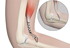
Elbow Ligament Reconstruction
Ligament reconstruction is considered in patients with ligament rupture. Your surgeon will make an incision over the elbow. Care is taken to move muscles, tendons, and nerves out of the way. The donor's tendon is harvested from either the forearm or below the knee.
Elbow Tendon and Ligament Repair
The common conditions affecting the tendons around the elbow joint include tennis elbow and golfer’s elbow, which result from an overuse injury to the tendons in weight lifting, or from repetitive activities during sports or occupation.
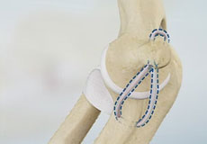
UCL Reconstruction (Tommy John Surgery)
Commonly called Tommy John surgery, this procedure involves reconstructing a damaged ligament on the inside of the elbow called the ulnar or medial collateral ligament with a tendon graft obtained from your own body or a donor.

Distal Biceps Repair
The biceps is a large muscle located in the front of your upper arm and runs from the shoulder to the elbow joint. It is attached to the bones of the shoulder and elbow by tendons. The distal biceps is the area where the biceps is attached to the forearm bone in the elbow.
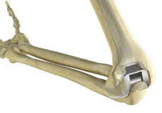
Total Elbow Replacement
Elbow joint replacement, also referred to as total elbow arthroplasty, is an operative procedure to treat the symptoms of arthritis that have not responded to non-surgical treatments. The goal of elbow joint replacement surgery is to eliminate your pain and increase the mobility of your elbow joint.
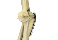
Elbow Fracture Reconstruction
Elbow fracture reconstruction is a surgical procedure employed to repair and restore the appearance and full function of a damaged elbow caused by severe trauma or injury. This may include repairing damaged structures or replacing missing or damaged structures with adjoining skin, muscles, ligaments, tendons, bones, or nerves to restore the appearance and function.
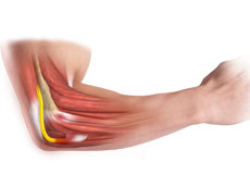
Tennis Elbow
Tennis elbow is a painful condition occurring from repeated muscle contractions at the forearm. The condition is more common in sports activities such as tennis, painting, hammering, typing, gardening and playing musical instruments.
Golfer's Elbow
Golfer’s elbow, also called medial epicondylitis, is a painful condition occurring from repeated muscle contractions in the forearm that leads to inflammation and microtears in the tendons that attach to the medial epicondyle.
Ulnar Nerve Entrapment at the Elbow (Cubital Tunnel Syndrome)
When the elbow is bent, the ulnar nerve can stretch and catch on the bony bump. When the ulnar nerve is compressed or entrapped, the nerve can tear and become inflamed, leading to cubital tunnel syndrome.
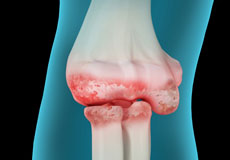
Elbow Arthritis
Although the elbows are not weight-bearing joints, they are considered to be most important for the functioning of the upper limbs. Hence, even minor trauma or disease affecting the elbow may cause pain and limit the movements of the upper limbs. Arthritis is one of the common disease conditions affecting the elbow joint.
Elbow (Olecranon) Bursitis
Inflammation of the olecranon bursa leads to a condition called olecranon bursitis.
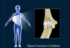
Elbow Fracture
Elbow fractures may occur from trauma, resulting from various reasons: a fall on an outstretched arm, a direct blow to the elbow or an abnormal twist to the joint beyond its functional limit.
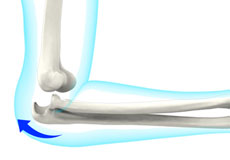
Elbow Dislocation
The arm in the human body is made up of three bones that join to form a hinge joint called the elbow. The upper arm bone or humerus connects from the shoulder to the elbow to form the top of the hinge joint. The lower arm or forearm consists of two bones, the radius, and the ulna. These bones connect the wrist to the elbow forming the bottom portion of the hinge joint.
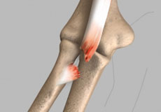
Bicep Tendon Tear at the Elbow
A biceps tear can be complete or partial. Partial biceps tendon tears will not completely break the tendon while complete tendon tears will break the tendon into two parts. Tears of the distal biceps tendon are usually complete and the muscle is separated from the bone. Tears of the distal biceps tendon most often result from a sudden injury or lifting a heavy object.
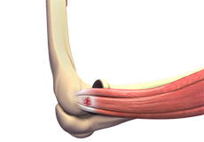
Elbow Anatomy
The elbow is a complex joint formed by the articulation of three bones – the humerus, radius, and ulna. The elbow joint helps in bending or straightening of the arm to 180 degrees and lifting or moving objects.
The bones of the elbow are supported by:
- Ligaments and tendons
- Muscles
- Nerves
- Blood vessels
Bones and Joints of the Elbow
The elbow joint is formed at the junction of three bones:
- The humerus (upper arm bone) forms the upper portion of the joint. The lower end of the humerus divides into two bony protrusions known as the medial and lateral epicondyles, which can be felt on either side of the elbow joint.
- The ulna is the larger bone of the forearm located on the inner surface of the joint. It articulates with the humerus.
- The radius is the smaller bone of the forearm situated on the outer surface of the joint. The head of the radius is circular and hollow, which allows movement with the humerus. The articulation between the ulna and radius helps the forearm to rotate.
The elbow consists of three joints, namely:
- The humeroulnar joint is formed between the humerus and ulna and allows flexion and extension of the arm.
- The humeroradial joint is formed between the radius and humerus and allows movements like flexion, extension, supination, and pronation.
- The radioulnar joint is formed between the ulna and radius bones and allows rotation of the lower arm.
Articular cartilage lines the articulating regions of the humerus, radius, and ulna. It is a thin, tough, flexible and slippery surface that acts as a shock absorber and cushion to reduce friction between the bones. The cartilage is lubricated with synovial fluid, which further enables the smooth movement of the bones.
Muscles of the Elbow Joint
There are several muscles extending across the elbow joint that help in various movements. These include the following:
- Biceps brachii: Upper arm muscle, enabling flexion of the arm
- Triceps brachii: Muscle in the back of the upper arm that extends the arm and fixes the elbow during fine movements
- Brachialis: Upper arm muscle beneath the biceps, which flexes the elbow towards the body
- Brachioradialis: Forearm muscle that flexes, straightens and pulls the arm at the elbow
- Pronator teres: Muscle that extends from the humeral head, across the elbow, and towards the ulna, and helps to turn the palm facing backward
- Extensor carpi radialis brevis: Forearm muscle that helps in movement of the hand
- Extensor digitorum: Forearm muscle that helps in movement of the fingers
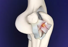
Ligaments and Tendons of the Elbow
The elbow joint is supported by ligaments and tendons, which provide stability to the joint.
Ligaments are a group of firm tissues that connect bones to other bones. The most important ligaments of the elbow joint are the:
- Medial or ulnar collateral ligament: Comprised of triangular bands of tissue on the inner side of the elbow joint
- Lateral or radial collateral ligament: A thin band of tissue on the outer side of the elbow joint
- Annular ligament: Group of fibers that surround the radial head, and hold the ulna and radius tightly in place during movement of the arm
Together, the medial and lateral ligaments are the main source of stability and hold the humerus and ulna tightly in place during movement of the arm.
The ligaments around a joint combine to form a joint capsule that contains synovial fluid.
Any injury to these ligaments can lead to instability of the elbow joint.
Tendons are bands of connective tissue fibers that connect muscle to bone. The various tendons that surround the elbow joint include:
- Biceps tendon: attaches the biceps muscle to the radius, allowing the elbow to bend
- Triceps tendon: attaches the triceps muscle to the ulna, allowing the elbow to straighten
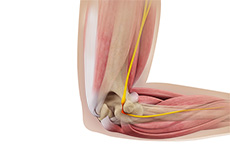
Nerves of the Elbow
The main nerves of the elbow joint are the ulnar, radial and median nerves. These nerves transfer signals from the brain to the muscles that aid in elbow movements. They also carry sensory signals such as touch, pain, and temperature back to the brain.
Any injury or damage to these nerves causes pain, weakness or joint instability.
Blood Vessels Supplying the Elbow
Arteries are blood vessels that carry oxygen-pure blood from the heart to the hand. The main artery of the elbow is the brachial artery that travels across the inside of the elbow and divides into two small branches below the elbow to form the ulnar and the radial artery.